‘Making Biology students interested in Etymologies’ !!!
· FUN TO LEARN BIOLOGICAL TERMINOLOGY
· THE LANGUAGE OF BIOLOGISTS
· SOWING SEEDS OF SYSTEMATICS / TAXONOMY AT THE GRASSROOT LEVEL
The Episode:
The episode of ‘Bio-etymology’ is devoted to analyzing the hidden meanings derived from the Names of various Animal Phyla and Classes, along with the terms specifically used to describe their respective diagnostics, important examples (Genus or species) etc.
Recollecting the Introduction of PART – 1:
At any level, may it be animals in general or Man in particular, there is some structured or indicative or behavioural system of communication. It is simply referred to as a kind of ‘Language’. In a broader sense, ‘Language’ is the method of communication that involves the use of various languages (in various countries) spoken by man. Articulation of words in a definite sequence is the basic of formulating a Language and knowledge of words forming it and their ‘sense’ is of utmost importance. Accumulation of a treasure of words constitutes what is called ‘Vocabulary’ defined variously as follows:
1. The words of a language.
2. The body of words used in a particular language.
3. All the words that exist in a particular language or subject.
4. A list or collection of the words or phrases of a language, technical field etc.
5. A listing either selective or exhaustive, containing the words and phrases of a language, with meaning or translations into another language.
Over a period of time in past centuries, Science is general and Biology in particular has accumulated a vast array of words to communicate fact(s) or phenomena through deriving their meanings.
Recollecting previous episodes of Bio-etymology
PART – 5
Division: BILATERIA
[Gk. bi = two + lateros = sides]
Diagnostics of Bilateria:
In contrast to Radiata [Phylum – Cnidaria; Bio-etymology PART –4], the remaining
multicellular animals belonging to BILATERIA have the following diagnostics in common viz., 1. Symmetry: ‘Bilateral’ [Gk. bi = two + lateros = sides] i.e., body divisible
into two identical halves (right and left) only in one plane passing through the median longitudinal axis.
2. Number of Germ Layers: Three (3) i.e., Triploblastic [Gk. triploos / triples;
Latin triplus = threefold / three + blast (os) = denoting embryonic cell / germ layer of an embryo / germ / sprout + -ic < Latin –icus / Gk. –ikos = the suffix used to form adjectives] i.e., the animal body developing from three primary germ layers viz., Ectoderm, Endoderm and Mesoderm.
3. Body Organization: Organ-system grade, with various tissues organizing into distinct
organs and organs into systems.
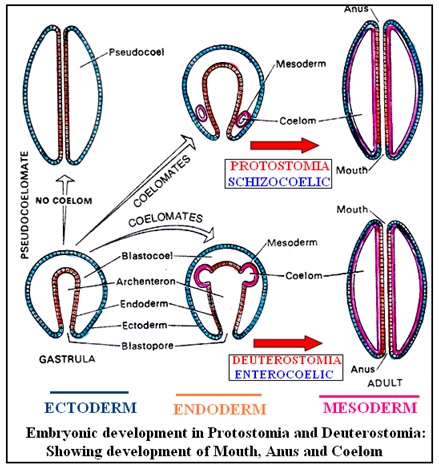
4. Development of mouth in the embryo:
There are two situations noticed viz., mouth of the adult develops from or close to the ‘blastopore’ (= Protostomia condition) or mouth develops opposite to the blastoporal end or blastopore becomes anus (= Deutrostomia condition).
5. Cephalization [Gk kephale = head, uppermost or top part]: The bilateral symmetry is correlated with the locomotor movements and one end of the body having the mouth always faces forward direction, the anterior. There is also a great concentration of nervous system and sense organs (eyes, ears etc.) on this anterior end, called the head. Cephalization, is thus, “a tendency to evolve in manner so as to have important parts near the head” (James D. Dana, 1813 – 1895, US Zoologist).
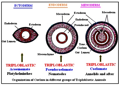
6. A ‘true body cavity’ or COELOM [Gk. koiloma / koilia = hollow, cavity]: Present
or absent.
· ‘Body cavity’: When interpreting a ‘body cavity’ it simply communicates about any internal space or a series of spaces present inside the body e.g., a cavity (or cavities) inside
a Sponge or a Cnidarian.
· A ‘true body cavity’ or COELOM: This refers to a large fluid-filled space (cavity) lying between the outer body wall and the inner digestive tube; arising as a secondary cavity between two layers of embryonic mesoderm or coelomic *epithelium [Gk. epi = above, upon + thele = teat, nipple] or **peritoneum
*Epithelium: This term was originally used to describe a translucent covering of small ‘nipples’. In 1703, a Dutch Botanist and anatomist, Fredrik Ruysch (1638 – 1731) used the term ‘Epithelia’ to describe the type of tissue or the translucent covering of small ‘nipples’, while dissecting the lip of a ‘cadaver’(= a dead human body used by medical students). In modern sciences, an ‘epithelium’ is a type of animal tissue, where the cells are packed into sheets that line internal surfaces and cover the external surface of the body. **Peritoneum: [< Latin peritonaeum / Gk. peritonaios = membrane lining the abdominal cavity; < Gk. peritonaion < peritonos = stretched over] [< Gk. peri = around + teinein / tonos = to stretch or stretched i.e., a membrane stretched over]: A thin mesodermal membrane lining the †abdominal and pelvic cavities and most of the ††abdominal viscera; hence of two types viz., † Parietal Peritoneum or Somatic Mesoderm: [Latin parietalis / paries / pariet = wall or pertaining to the walls of a cavity] [Gk. somatikos / soma = of the body / the body] i.e., the outer mesodermal layer present just below the body wall (= ectoderm). †† Visceral Peritoneum or Splanchnic Mesoderm: [< Latin visceralis / viscera = internal organ, inner parts of body] [Gk. splankhon = pertaining to viscera] i.e., the mesodermal layer covering all the internal (visceral) organs. |
TYPES OF COELOM
Based on the mode of differentiation, Primary and Secondary are the two types of Coeloms:
Primary Coelom or Pseudocoelom [Gk. pseudo = false, feigned, erroneous + coelom]:
This kind of ‘Coelom’ is called ‘false coelom’ because it is a cavity between ectoderm on the outside and endoderm (digestive tract) inside [i.e., not lined by mesoderm on both the sides]. In fact, this kind of body cavity is derived from the ‘blastocoel’ of the embryo, rather representing the persistent blastocoel.
Examples: Nematodes (Round Worms).
Secondary Coelom:
In all the higher bilateria, from Annelids onwards, the blastocoel gets obliterated by the development of endodermal ‘archenteron’ [embryonic gut] and another space (true coelom or eucoelom) is created between two layers of mesodermal cells (peritoneum) present between the gut (endoderm) and the body wall (ectoderm).
Examples: Annelida to Chordata.
7. Metamerism (Segmentation):
[Gk. meta = after + meros = a part, a fraction]
[Segment + -ation < Latin segmentum / Gk. tmema or tmemata = a strip or piece cut off; a division, section + -ation = a suffix used in nouns, denoting ‘action’]
Body of bilaterally symmetrical animals may or may not be divided into a linear series of parts (called segments), a phenomenon being called as ‘metamerism’. This segmentation may further be ‘Pseuso– (false)’ or ‘true’:
Pseudometamerism vs True Metamerism
· Pseudometamerism refers only to the superficial segmentation or repetition of similar, complete, reproductive individuals (functionally independent); the youngest one being added from the front (anterior end). This kind of segmentation is also called ‘strobilation’ [Gk. strobile / strobilus = twisted plug of lint, shaped like a ‘pine cone’] and each segment is named as proglottid [Gk. proglossis / proglossid = point / tip of the tongue; based on glossa / glotta = tongue; pro = before + glossa / glotta = tongue] because of their shape. Thus, there is serial repetition of ‘proglottids’, each containing a complete set of reproductive organs (independent of the others); as exemplified by ‘Tapeworms’ (Platyhelminthes).
· True metamerism is what is defined ProtostomeAnnelida, Mollusca and Arthropoda) and Deutrostome
Eucoelomates (Echinodermata,
Hemichordata and Chordata )
where there is a longitudinal division of body into a linear
series of similar sections / parts,
called the metameres or segments and the phenomenon being
called as ‘metamerism’.
In this case the number of segments is fixed, functionally interdependent and
laid down in the embryonic stages, the youngest one being the posteriormost.
Each metamere also typically repeats some or all of the various organ units.
The primary segmental divisions are – body wall, musculature and often the
coelom.
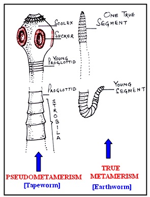
Annelids exhibit both external and internal metamerism whereas in Arthropods it is chiefly external but in man and other Vertebrates it is only internal metamerism. Therefore, the segmentation is again of two types:
· Homonomous or Complete Metamerism [Gk. homos = same + nomos = law]:
In this kind of metamerism, the metameres / segments are all alike (= homonomous); affecting practically all the systems (= complete) i.e., there are segmental blood vessels, nerves, nephridia, gonads, as exemplified by Annelids.
· Heteronomous or Incomplete Metamerism [Gk. heteros = different, other + nomos = law]:
In post-larval/embryonic development of Arthropods and Vertebrates, on account of specialization or modification, the metameres / segments of different regions become greatly dissimilar e.g., more simplification by loss of segments, fusion of segments (cephalization), disappearance of certain structures or appearance of other structures (like limbs) etc. Hence, such a metamerism is called incomplete or heteronomous.
SUBDIVISIONS OF BILATERIA
On the basis of the embryonic development and the fate of the ‘blastopore’ being decided into either ‘mouth’ or ‘anus’, the bilaterally symmetrical animals are placed under two Subdivisions:
[A] SUBDIVISION – PROTOSTOMIA: [Gk. protos = first + stoma = mouth] i.e., the ‘mouth’ of the adult develops from the ‘first mouth’ of the embryo, the Blastopore (of Gastrula) or close to the Blastopore. This term was coined by an Austrian Biologist, Karl Grobben (1854 – 1945).
[B] SUBDIVISION – DEUTEROSTOMIA: [Gk. deuteros = second, next + stoma = mouth] i.e., the ‘mouth’ of the adult develops away from or opposite the ‘first mouth’ of the embryo, the Blastopore (in Gastrula stage) [the Blastopore becoming the anus]. This term was coined by an Austrian Biologist, Karl Grobben (1854 – 1945).
SUBDIVISION – PROTOSTOMIA
Based on the presence or absence of a ‘true coelom’, Protostomes are classified under 3 SECTIONS:
[A] Acoelomata [Gk. a = no, without + koiloma / koilia = hollow, cavity + ata = a group or a group of organisms characterized by a structure]:
There is no coelom, as embryonic mesoderm remains as a solid mass (layer) and the space between the endoderm (gut wall) and the ectoderm (body wall) remains filled with mesodermal tissue called mesenchyme [Gk. mesos = middle + enkhyma = infusion] i.e., a layer infused in the middle; often called ‘packing tissue’ or parenchyma [Gk. para = beside + enkhyma = infusion i.e., something poured in beside].
Examples: Phylum – Platyhelminthes (Flatworms) and Nimertinea (Ribbonworms).
[Bio-etymology PART – 6 & 7 ]
[B] Pseudocoelomata [Gk. pseudo = false + koiloma / koilia = hollow, cavity + ata = a group or a group of organisms characterized by a structure]:
A ‘false coelom’ or a cavity between ectoderm on the outside and endoderm (digestive tract) inside [i.e., not lined by mesoderm on both the sides]. In fact, this kind of body cavity is derived from the ‘blastocoel’ of the embryo, rather representing the persistent blastocoel.
Example: Phylum – Nematoda (Round worms).
[Bio-etymology PART – 8]
[C] Eucoelomata [Gk. eu = true + koiloma / koilia = hollow, cavity + ata = a group or a group of organisms characterized by a structure]:
The ‘Coelom’ is a space enclosed by mesoderm on both sides or true coelom or eucoelom is created between two layers of mesodermal cells (peritoneum) present between the gut (endoderm) and the body wall (ectoderm).
On the basis of embryonic development, the Coelom of Protostomes is of chizocoelous type or they exhibit ‘Schizocoely’ [Gk. skhizo = to split + koiloma / koilia = hollow, cavity] i.e., coelom arises by a splitting of endomesodermal cells which originate from the blastoporal region of the larva and extend between ectoderm and mesoderm.
Examples: From Phylum – Annelida, Mollusca and Arthropoda.
[Bio-etymology PART – 9, 10 & 11]
SUBDIVISION – DEUTEROSTOMIA
Based on the development of ‘true coelom’, all Deuterostomes are ‘Eucoelomates’ and it is of Enterocoelous type or they exhibit ‘Enterocoely’ [Gk. enteron = gut / intestine + koiloma / koilia = hollow, cavity] i.e., coelom arises as the mesodermal pouches from larval archenteron (embryonic gut).
Examples: Echinodermata, Hemichordata and Chordata.
[Bio-etymology PART – 12 ,13 & 14]
|
Learning
process is an on-going process: Keep
on venturing more into the fantastic world of Etymology and feel Bilateria –
friendly!!! |